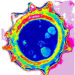
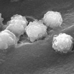
Transmission electron micrograph of a C. neoformans

Date: 3 April 2014

Transmission electron micrograph of a C. neoformans cell seen in CSF in an AIDS patients with remarkably little capsule present. These cells may be mistaken for lymphocytes.

Copyright: n/a

Notes: n/a


Images library

Showing 10 posts of 2574 posts found.
  • Title

    Legend

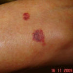
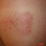
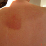
  • Drug rashes: Drug interactions between steroids and anti-fungal drugs – (ecchymosis)
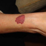
    Image A: Red skin rash on arms due to effect of inhaled steroids termed ecchymosis., Image B: Red skin rash on arms due to effect of inhaled steroids termed ecchymosis., Image C: Papular rash with blisters on the back, associated with CPA but prior to treatment, rash appeared 2 years previously in October 2007. The rash resolved on treatment with azoles., Image D: Papular rash with blisters on the back, associated with CPA but prior to treatment, rash appeared 2 years previously in October 2007. The rash resolved on treatment with azoles.

  • Reference: Muco-cutaneous retinoid effects and facial erythema related to the novel triazole antifungal agent voriconazole. Denning, DW & Griffiths, CEM. Clin.Exp Dermatol 2001, 26(8), 648-53.
    Courtesy of Dr D Denning, Wythenshawe Hospital, Manchester.(© Fungal Research Trust)
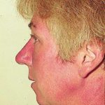
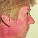
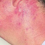
    Patient AB: Voriconazole rash. Following 8 weeks of Voriconazole, patient had remarkable facial erythema, most consistent with sun exposure because of sparing of her neck., Patient AB: Voriconazole rash. Following 8 weeks of Voriconazole, patient had remarkable facial erythema, most consistent with sun exposure because of sparing of her neck., Patient AB: Discoid lupus erythematosus following 12 months voriconazole therapy. This improved with use of sunblock factor 30 and resolved after discontinuation of voriconazole, 2 months later.

  • Micrographs of A. niger conidia & conidial heads provided by Amaliya Stepanova, Head of Laboratory pathomorphology and cytology at Kashkin Research Institute, Russian Federation.
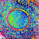
    A niger conidial head TEM in vitro (РКПГF-1124), A niger mature conidium TEM in vitro (РКПГF-1124)

  • Micrographs of A. terreus conidia & conidial heads provided by Amaliya Stepanova, , Head of Laboratory pathomorphology and cytology at Kashkin Research Institute, Russian Federation.
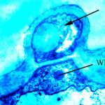
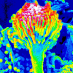
    A terreus aleurospore TEM in vitro (РКПГF-1275), A terreus mature conidium SEM in vitro (РКПГ-1275), A terreus mature conidial head SEM in vitro

  • Micrographs of A. fumigatus conidia & conidial heads provided by Amaliya Stepanova, , Head of Laboratory pathomorphology and cytology at Kashkin Research Institute, Russian Federation.
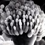
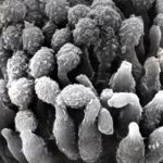
    Conidial head (SEM), Part of conidial head (SEM), Mature conidia (SEM), Hyphae (SEM), Murine lung tissue (TEM)

  • Isolate from environmental sample

    A. clavatus, A. clavatus, A. clavatus

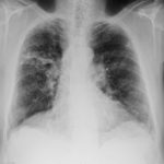
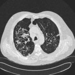
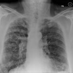
  • Patients has history of ABPA complicating long standing asthma. His total IgE has fluctuated between 2,200 and 4,600 KU/L, his Aspergillus IgE between 36.3 and 65.4 kAU/L and Aspergillus IgG from 87-154 mg/L. He has been taking long term itraconazole.
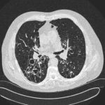
    December 2012, May 2012, AW CT Dec 2012 2, February 2010