Date: 26 November 2013
Secondary metabolites, 3D structure: Trivial name – Xanthone derived
Copyright: n/a
Notes:
Species: A. wentiiSystematic name: 9H-Xanthene-1-carboxylic acid, 3,8-dihydroxy-6-methyl-9-oxo-, methyl esterMolecular formulae: C16H12O6Molecular weight: 300Chemical abstracts number: 85003-85-6Selected references: Isolation and structures of four new metabolites from Aspergillus wentii. Hamasaki, Takashi; Kimura, Yasuo. Dep. Agric. Chem., Tottori Univ., Tottori, Japan. Agricultural and Biological Chemistry (1983), 47(1), 163-5. CODEN: ABCHA6 ISSN: 0002-1369. Journal written in English. CAN 98:122499 AN 1983:122499 CAPLUS
Images library
-
Title
Legend
-
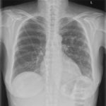
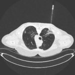
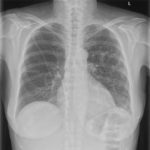
PtDS2 –Repeated chest infections arrested by itraconazole therapy in ABPA and bronchiectasis
DS2 developed asthma age 24 and now aged 62. From about age 30 she started getting repeated chest infections and a few years later ABPA and bronchiectasis was diagnosed. Infections continued requiring multiple courses of antibiotics annually. At one point DS2 developed a pneumothorax, possibly because of excess coughing. She has chronic rhinitis and mannose binding lectin deficiency. In May 2011, she started itraconazole therapy, and has needed no antibiotic courses for her chest since. Her rhinitis with sinusitis occasionally bothers her. She is delighted to have gone 18 months with no chest infections.
 ,
,  ,
, 
-
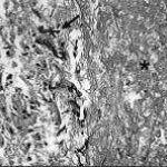
Aspergillus hyphae (arrow) in the lumen without invasion of the necrotic bronchial wall (*) (Nicod 2001).
-
fibrinonecrotic material (arrow) from the airway shown in A, with subocclusion of the bronchial lumen (*)
-
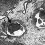
Fibrinous or pseudomembranous bronchitis (arrow) with subocclusion of the airways (* indicates subocclusion of the airways by pseudomembranes)

-
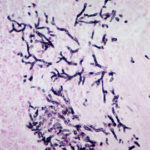
Bronchoscopic biopsy demonstrated septate hyphae with branching at 45o (methenamine silver stain ×400).
