Date: 26 November 2013
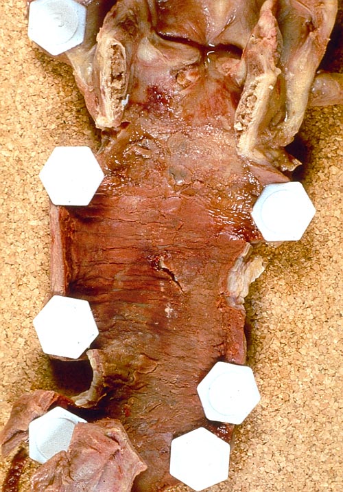
Pt FT. Autopsy appearance of the trachea, after the adherent pseudomembrane had been removed, revealing confluent ulceration superiorly with small green plaques of Aspergillus growth on the trachea inferiorly.
Copyright: n/a
Notes:
This patient had an acute onset of neutropenia, of undetermined origin, which was treated with prednisolone, before developing rapidly progressive and ultimately fatal pseudomembranous Aspergillus tracheobronchitis. His case was reported because he developed a unilateral monophonic wheeze, which prompted a diagnostic bronchoscopy.Tait RC, O’Driscoll BR, Denning DW. Unilateral wheeze due to pseudomembranous Aspergillus tracheobronchitis in the immunocompromised patient. Thorax 1993; 48: 1285-1287. Disseminated aspergillosis was found at autopsy and cultures from each organ were found to be clonal (Birch M, Nolard N, Shankland G, Denning DW. DNA typing of epidemiologically-related isolates of Aspergillus fumigatus. Infect Epidemiol 1995; 114: 161-168.)
Images library
-
Title
Legend
-
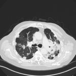
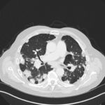
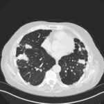
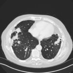
Mr RM is 80 and an ex-coal miner.He developed pneumoconiosis from exposure to coal dust. He also developed rheumatoid arthritis and the combination of this disease and pneumoconiosis is called Caplan’s syndrome.
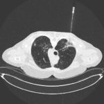
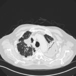
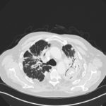
His chest Xray in early 2015 shows extensive bilateral pulmonary shadowing with solid looking nodules superimposed on abnormal lung fields, contraction of his left lung with an elevated diaphragm and a large left upper lobe aspergilloma, displaying a classic air crescent. His CT scan from mid 2014 demonstrates a large aspergilloma in a cavity on the left, with marked pleural thickening around it, which is partially ‘calcified’ towards its base. Inferiorly on other images,remarkable pleural thickening and fibrotic irregular and spiculated nodules are seen, most partially calcified.
 ,
,  ,
,  ,
,  ,
,  ,
,  ,
, 