Date: 21 January 2014
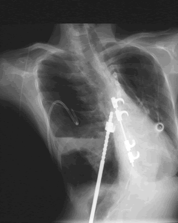
The chest is distorted by a deformity of the back and ribs.
Copyright: n/a
Notes:
This patient’s X-ray is complex. The chest is distorted by a deformity of the back and ribs. Substantial metalwork following a spinal fusion is in place to support the vertebral column and part of this overlies the heart and part of it crosses the left lung. The patient also has a portacath device in-situ over the right lung, which allows i.v. antibiotics to be given. A needle is in-situ inside the portacath device. An external drainage tube is currently in-situ in a large air cavity and left upper thorax. This cavity contains mostly air but there is some fluid with the fluid level at its base. Underneath this large pyopneumothorax is a normal component of left lower lobe. The heart is very substantially moved to the right of the lung because of a previous right lower lobe resection. There is no evidence of aspergillosis on this x-ray as it stands.
Images library
-
Title
Legend
-
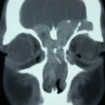
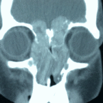
4 Total obstruction of the sinuses due to inflamed mucosa. (Patient 04)
-
1 Axial computed tomography (CT) scans of the frontal sinus.
A: due to the long lasting pressure of mucus, the bone of the anterior wall of frontal sinus is thinned out and elevated anteriorly, forming a bulge. B: same situation as depicted in fig A: the posterior bony wall of frontal sinus is thinned out and extremely elevated posteriorly towards the frontal lobe of the brain. As depicted on the scan, a thin bony layer covering the dura could be recognized intraoperatively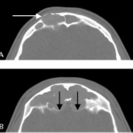
-
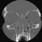
2 Same patient as 1 and 3, frontal CT
-
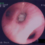
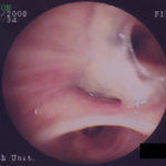
D. 6 months later, tenacious yellow secretions in L basal bronchial division
-
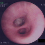
C. After suction the material was seen to extend distally – obstructing the right basal stem bronchus
-
B. After suction the material was seen to extend distally – obstructing the right basal stem bronchus
