Date: 21 January 2014
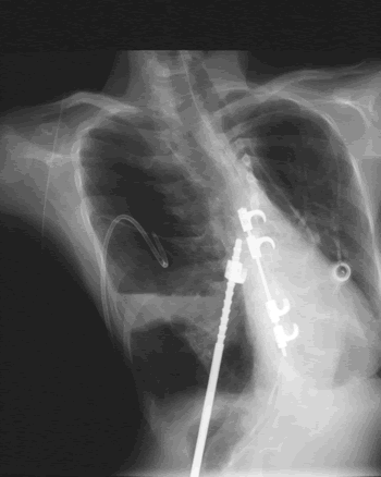
The chest is distorted by a deformity of the back and ribs.
Copyright: n/a
Notes:
This patient’s X-ray is complex. The chest is distorted by a deformity of the back and ribs. Substantial metalwork following a spinal fusion is in place to support the vertebral column and part of this overlies the heart and part of it crosses the left lung. The patient also has a portacath device in-situ over the right lung, which allows i.v. antibiotics to be given. A needle is in-situ inside the portacath device. An external drainage tube is currently in-situ in a large air cavity and left upper thorax. This cavity contains mostly air but there is some fluid with the fluid level at its base. Underneath this large pyopneumothorax is a normal component of left lower lobe. The heart is very substantially moved to the right of the lung because of a previous right lower lobe resection. There is no evidence of aspergillosis on this x-ray as it stands.
Images library
-
Title
Legend
-
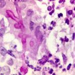
BAL specimen showing hyaline, septate hyphae consistent with Aspergillus, stained with Blankophor
-
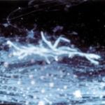
Mucous plug examined by light microscopy with KOH, showing a network of hyaline branching hyphae typical of Aspergillus, from a patient with ABPA.
-
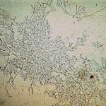
Corneal scraping stained with lactophenol cotton blue showing beaded septate hyphae not typical of either Fusarium spp or Aspergillus spp, being more consistent with a dematiceous (ie brown coloured) fungus
-
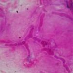
Corneal scrape with lactophenol cotton blue shows separate hyphae with Fusarium spp or Aspergillus spp.
-
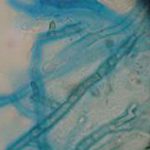
A filamentous fungus in the CSF of a patient with meningitis that grew Candida albicans in culture subsequently.
-
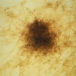
Transmission electron micrograph of a C. neoformans cell seen in CSF in an AIDS patients with remarkably little capsule present. These cells may be mistaken for lymphocytes.
-
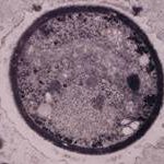
India ink preparation of CSF showing multiple yeasts with large capsules, and narrow buds to smaller daughter cells, typical of C. neoformans
