Date: 3 April 2014
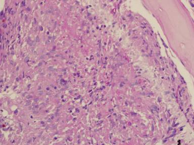
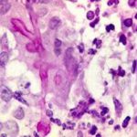
PAS stain. An example of Aspergillus fumigatus.
(PAS-stained) in a patient with chronic granulomatous disease showing a 45 degree branching hypha within a giant cell. Rather bulbous hyphal ends are also seem, which is sometimes found inAspergillus spp. infections, histologically. (x800)
Copyright: n/a
Notes:
Comparison of GMS and PAS stains. Patient with disseminated Trichosporon spp. infection. Both x60. In the GMS image, substantial background staining of elastin is seen, with more prominent yeasts superimposed. In contrast, the PAS stain shows the tissue morphology, with bright pink yeasts also visible.
Images library
-
Title
Legend
-
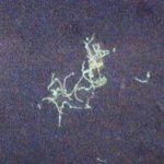
BAL specimen showing hyaline, septate hyphae consistent with Aspergillus, stained with Blankophor
-
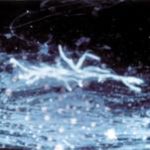
Mucous plug examined by light microscopy with KOH, showing a network of hyaline branching hyphae typical of Aspergillus, from a patient with ABPA.
-
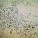
Corneal scraping stained with lactophenol cotton blue showing beaded septate hyphae not typical of either Fusarium spp or Aspergillus spp, being more consistent with a dematiceous (ie brown coloured) fungus
-
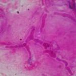
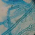
Corneal scrape with lactophenol cotton blue shows separate hyphae with Fusarium spp or Aspergillus spp.
-
A filamentous fungus in the CSF of a patient with meningitis that grew Candida albicans in culture subsequently.
-
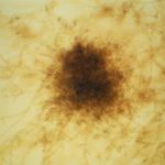
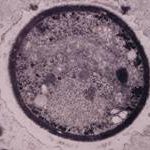
Transmission electron micrograph of a C. neoformans cell seen in CSF in an AIDS patients with remarkably little capsule present. These cells may be mistaken for lymphocytes.
-
India ink preparation of CSF showing multiple yeasts with large capsules, and narrow buds to smaller daughter cells, typical of C. neoformans
