Date: 6 August 2015
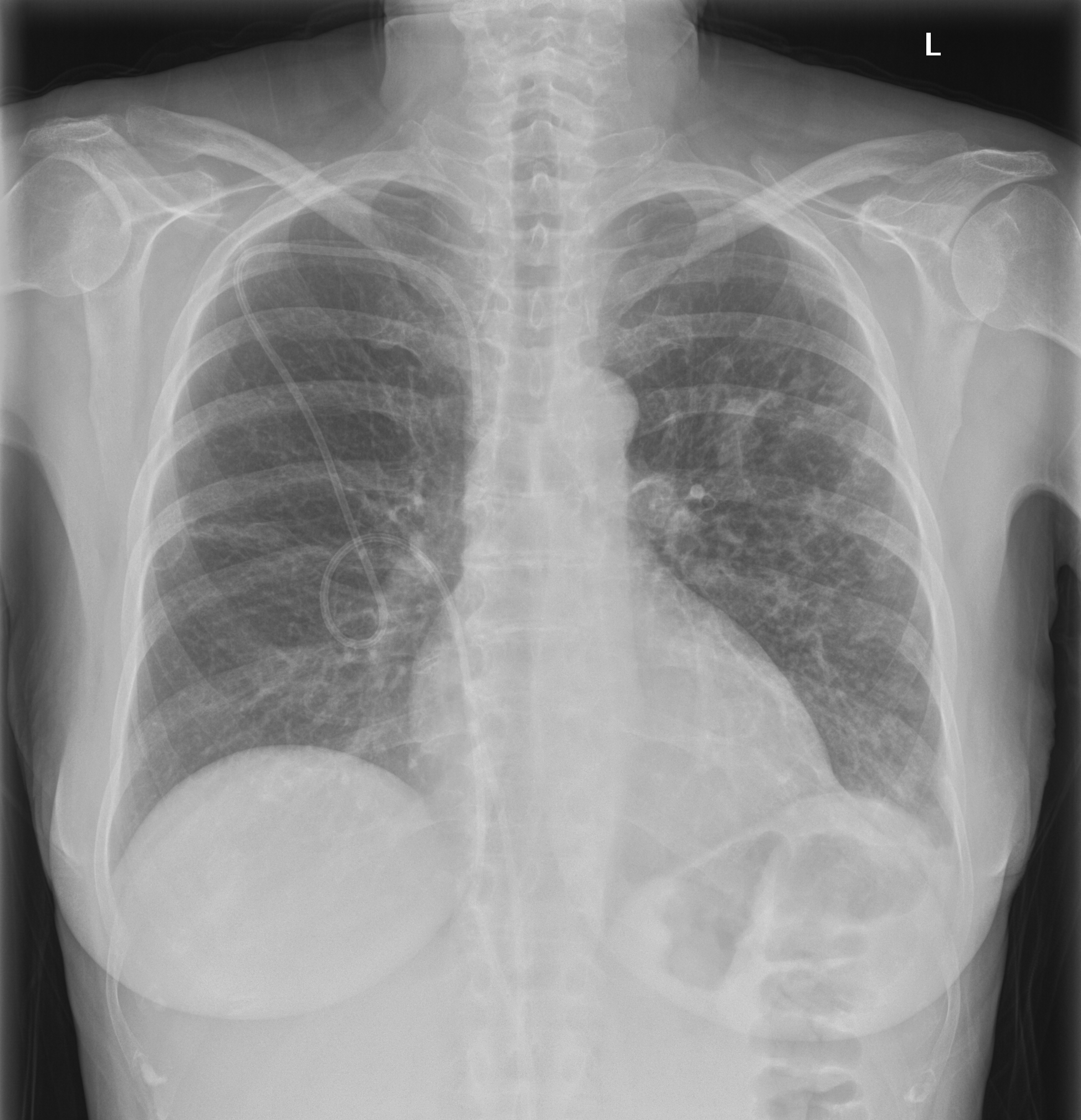
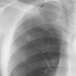
Chest Xray showing the normal course of a Hickman line
Copyright: n/a
Notes:
Chest Xray showing the normal course of a Hickman line, usually used for delivering intravenous medication, and taking blood, in leukaemia patients. The line as shown is partly in the body and partly over the skin of the chest. The Hickman line is placed just below the clavicle (collar bone) on the patients right side (it can be on the left) into the subclavian vein. It is then fed through to the superior vena cava which drains blood from the upper body, head and neck into the heart. The end of the line lies in the superior vena cava about 6 inches (15 cm) above the heart (right atrium).
Images library
-
Title
Legend
-
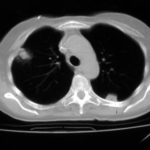
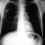
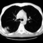
Nodules and areas of atelectasis are seen at both bases. He later died.

-
Further details
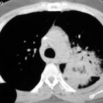
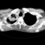
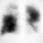
It is clearly a relatively small cavitary lesion, and the patient was almost asymptomatic. This response was a ‘stable’ response. The patient was included in the report Denning DW, Lee JY, Hostetler JS, Pappas P, Kauffman CA, Dewsnup DH, Galgiani JN, Graybill JR, Sugar AM, Catanzaro A, Gallis H, Perfect JR, Dockery B, Dismukes WE, Stevens DA, NIAID Mycoses Study Group multicenter trial of oral itraconazole therapy of invasive aspergillosis. Am J Med 1994; 97: 135-144.
 ,
, 
-
Well demarcated pulmonary infarction is well seen in this close-up of the lung at autopsy in a patient with histologically confirmed invasive aspergillosis. Angio invasion is characteristic of invasive aspergillosis, is associated with a worse prognosis, but is not always seen.

-
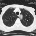
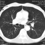
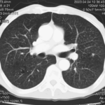
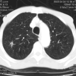
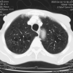
This 83 year old man presented with weight loss to a lung cancer clinic in mid 2003.
 ,
,  ,
,  ,
,  ,
,  ,
, 