Date: 6 August 2015
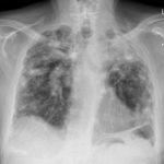
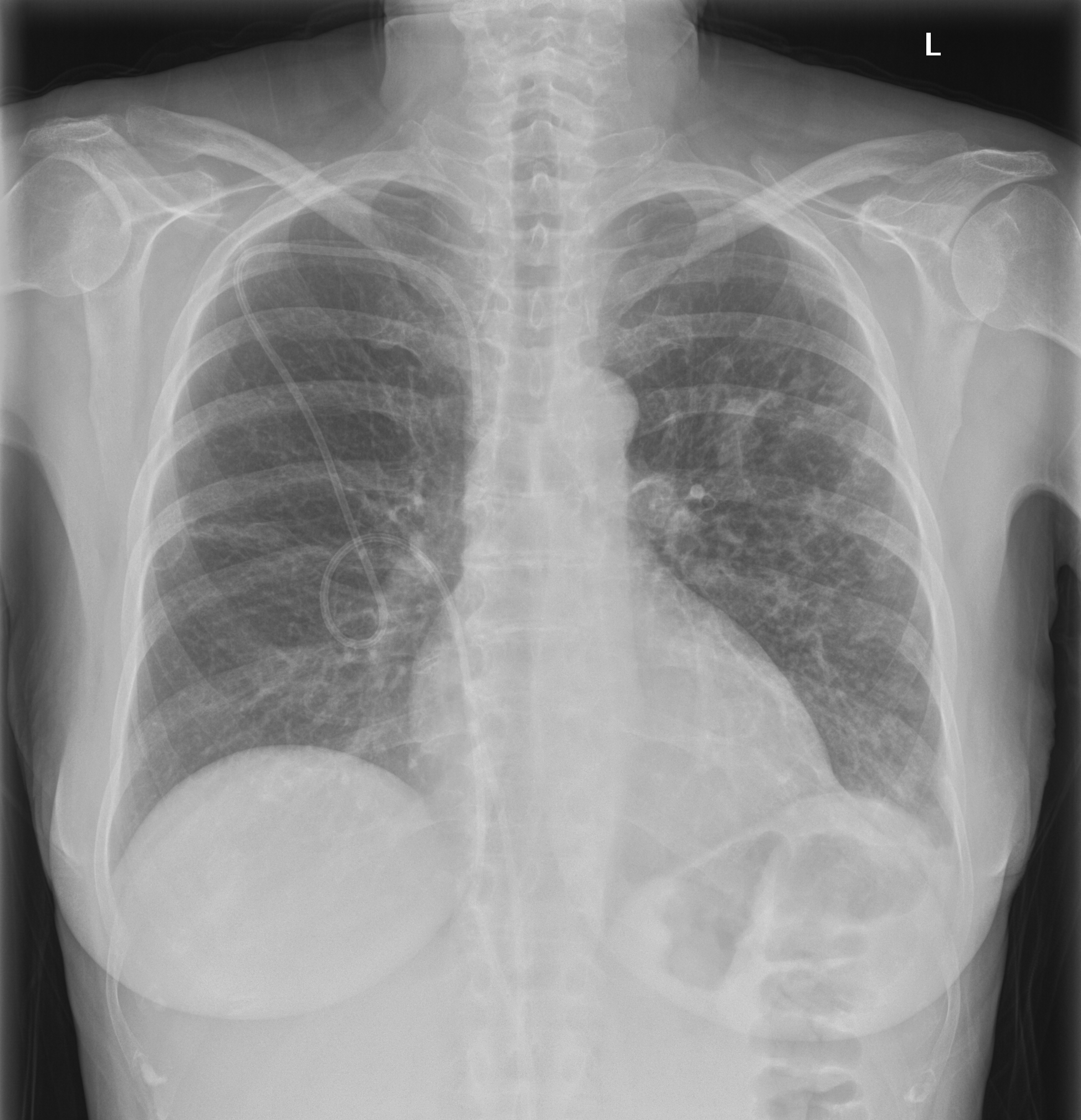
Chest Xray showing the normal course of a Hickman line
Copyright: n/a
Notes:
Chest Xray showing the normal course of a Hickman line, usually used for delivering intravenous medication, and taking blood, in leukaemia patients. The line as shown is partly in the body and partly over the skin of the chest. The Hickman line is placed just below the clavicle (collar bone) on the patients right side (it can be on the left) into the subclavian vein. It is then fed through to the superior vena cava which drains blood from the upper body, head and neck into the heart. The end of the line lies in the superior vena cava about 6 inches (15 cm) above the heart (right atrium).
Images library
-
Title
Legend
-
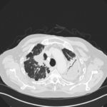
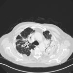
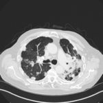
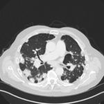
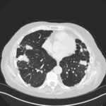
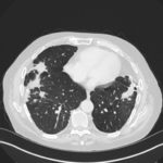
Mr RM is 80 and an ex-coal miner.He developed pneumoconiosis from exposure to coal dust. He also developed rheumatoid arthritis and the combination of this disease and pneumoconiosis is called Caplan’s syndrome.
His chest Xray in early 2015 shows extensive bilateral pulmonary shadowing with solid looking nodules superimposed on abnormal lung fields, contraction of his left lung with an elevated diaphragm and a large left upper lobe aspergilloma, displaying a classic air crescent. His CT scan from mid 2014 demonstrates a large aspergilloma in a cavity on the left, with marked pleural thickening around it, which is partially ‘calcified’ towards its base. Inferiorly on other images,remarkable pleural thickening and fibrotic irregular and spiculated nodules are seen, most partially calcified.
 ,
,  ,
,  ,
,  ,
,  ,
,  ,
, 