Date: 29 August 2017
Copyright:
Fungal Infection Trust
Notes:
ptAS is seen at the National Aspergillosis Centre in Manchester, UK and has ABPA and bronchiectasis
Images library
-
Title
Legend
-
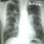
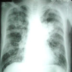
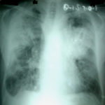
The chest X rays showed a rapid progression of lung disease- with bilateral upper zone and midzone consolidation and bilateral pleural effusion. Both lower lobes showed bronchiectasis in a central distribution along with centrilobular nodules and tree-in-bud pattern.
 ,
,  ,
,  ,
, 
-
Patient EG Intraluminal aspergilloma in cystic fibrosis (12 months follow up)
The 21 year old woman with cystic fibrosis developed an aspergilloma in her left lower lobe bronchus. CF was diagnosed at 6 months of age (sweat chloride 78 and 100 mmol/L) and CFTR mutations δ508 and W1282X and she developed diabetes mellitus at age 12 years. Age 15 years ABPA was diagnosed. Her serum IgE at the time of diagnosis was 5060 IU/L, skin prick test for aspergillus was positive, and serum was positive for precipitating antibodies to Aspergillus. She was treated with oral prednisone (1 mg/kg/day) for first two weeks followed by prednisone at 0.5 mg/kg every other day for at least 6 months with some clinical and serologic improvement. Over the following 5 years, she presented with a pattern of repeated episodic exacerbations with wheezing and crackles, increases in IgE and need to increase prednisone dosage.
In the 12 months before the aspergillomas were found, she started to experience frequent pulmonary exacerbations, which have prompted intensive therapies. She has also been on oral prednisone & itraconazole for at least 9 months for her ABPA relapse with some clinical & serologic improvement. She then developed severe protracted coughing spells associated with minor hemoptysis, low grade intermittent fever, and weight loss. Her FEV1 declined in a 3 months period from 56% to 33%. A recent chest-x ray did not reveal any new changes when compared to the one obtained almost a year before. A CT scan of the chest, however shows an ovoid soft tissue density within an ectatic bronchi in the anterior basal segment of the lower left lobe, felt to be an aspergilloma. [Link here].
She was started on voriconazole 200 mg twice daily. This dose gave a random serum level of 5ug/L. Her prednisone was weaned to 5 mgs/day and her FEV1 increased to 46% of predicted. In January 2009, her IgE level was 3053 kU/L; one year later, her IgE level was 1167 kU/L. A lung transplant surgeon attempted unsuccessfully to remove the aspergilloma via flexible bronchoscopy. It took a while for her to recover from that procedure. She then had a pulmonary exacerbation. She tolerated voriconazole reasonably well and gaining some weight. By mid 2010, her IgE level was 637 IU/L.
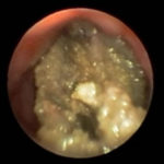
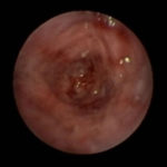
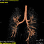
In early 2011, her aspergilloma (yellow friable material) in the anterior segment of the left lower lobe was removed completely (Fig a and b). It took a little time, but with biopsy forceps and some mincing and homogenization, it was all sucked out. The 3D reconstruction (Fig c) only shows the area of bronchiectasis, not the aspergilloma.Dr. Turcios is the director of pediatric pulmonology/cystic fibrosis in Somerville, NJ.
 ,
,  ,
, 
-
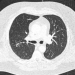
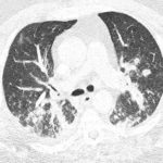
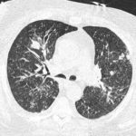
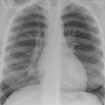
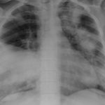
This patient is a 70 yr old, obese diabetic with aortic stenosis and COPD. He was admitted in early March 09, with collapse and loss of conciousness. His lungs appeared normal at this time – CT and X-ray 1. 10 days later he was admitted with increasing shortness of breath and chest X ray (F) showing widespread patchy consolidation. CT scan (B) showed bronchial dilatation, mucus plugging, nodular and bibasal consolidation. Multiple sputum samples grew Aspergillus fumigatus. The patient required intubation and remained in ITU for 160 days.
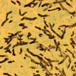
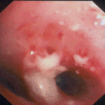
Bronchoscopy showed plaques in the major airways with more distal airways plugged with secretions resembling “cottage cheese”.There was severe contact bleeding and oedematous mucosa (I & J). Biopsy of the plaques showed fungal hyphae with a branching pattern consistent with aspergillus infection (G & H).
The patient was initially given IV and nebulised amphotericin B whilst on doses of hydrocortisone from 100-400 mg/day. Voriconazole was added with dose optimisation, and amphotericin discontinued. The patient improved gradually with voriconazole treatment over several months and for the latter month, gamma interferon was added into his regime which further improved his CT scan although some shadowing and bronchial wall thickening was still seen (D).
 ,
,  ,
,  ,
,  ,
,  ,
,  ,
,  ,
,  ,
,  ,
, 
-
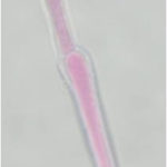
Hyphal septate club-like enlargements from culture on CYA 25°C medium (mag x100)
-
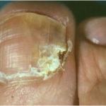
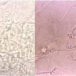
G Potassium hydroxide preparation of a nail specimen with onychomycosis- examined by microscopy
-
Culture plates on different media. A Colonies on CZ at 24oC, B on CYA at 20oC C on SAB at 37oC, D on CYA at 24oC


