Date: 26 November 2013
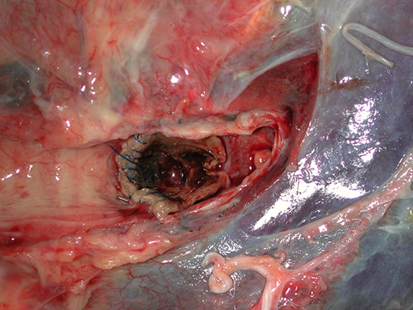
Fig2 Left main bronchus
Copyright: n/a
Notes:
Fig 1. Trachea and bronchi A 50+ year old woman received a double lung transplant for emphysema. She did well initially, but then Aspergillus fumigatus was grown from her airways, in association with mucous and a pseudomembrane covering parts of her anastomosis and airways. 2 months after her transplant she was undergoing bronchoscopy, and started to bleed. This rapidly became torrential and she suffered a cardiac arrest and died.
She underwent autopsy at which it was found that the larynx, trachea and major bronchi all contained blood (Fig 1).
The bronchial anastomoses were intact, but brown fluffy material was found overlying the stitches on both sides. On the right side plaques of similar material were seen distal to the anastomoses, overlying an ulcer and an obstructing the smaller bronchi.
On the left side an ulcer 1.5cm in diameter (with blood in it) was seen in the main bronchus distal to the anastomosis on the anterior wall (Fig 2). The sutures of the anastomosis are intact. The centre of the ulcer had ulcerated through into the left main pulmonary artery (Fig 3). The pulmonary artery shows necrosis and discolouration of the intimal surface over an area of 1.5-1.0cm.
Histopathology examination showed fungal hyphae perforating the bronchial wall and arterial wall around and in the ulcer. The ulcer on the right side showed hyphae perforating the wall and bronchial cartilage.
Images library
-
Title
Legend

