Date: 26 November 2013
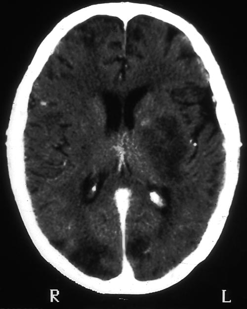
Contrast enhanced CT scan of the brain showing unequivocally 2 hypodense lesions, one in the left basal ganglia and one in the right occipital cortex. There is the possibility of another smaller left sided occiptal cortex. These lesions do not have the appearance of abscesses, but rather of ischaema.
Copyright: n/a
Notes: n/a
Images library
-
Title
Legend
-
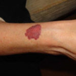
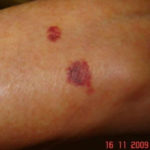
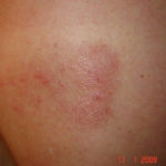
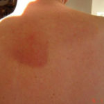
Drug rashes: Drug interactions between steroids and anti-fungal drugs – (ecchymosis)
 ,
,  ,
,  ,
, 
-
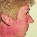
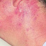
Reference: Muco-cutaneous retinoid effects and facial erythema related to the novel triazole antifungal agent voriconazole. Denning, DW & Griffiths, CEM. Clin.Exp Dermatol 2001, 26(8), 648-53.
Courtesy of Dr D Denning, Wythenshawe Hospital, Manchester.(© Fungal Research Trust)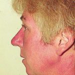 ,
,  ,
, 
-
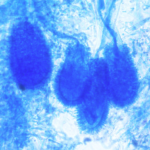
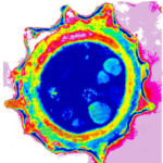
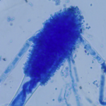
Micrographs of A. niger conidia & conidial heads provided by Amaliya Stepanova, Head of Laboratory pathomorphology and cytology at Kashkin Research Institute, Russian Federation.
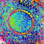 ,
, 
-
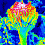
Micrographs of A. terreus conidia & conidial heads provided by Amaliya Stepanova, , Head of Laboratory pathomorphology and cytology at Kashkin Research Institute, Russian Federation.
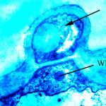 ,
,  ,
, 
-
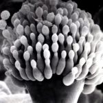
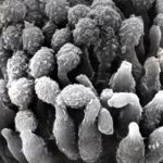
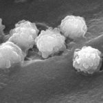
Micrographs of A. fumigatus conidia & conidial heads provided by Amaliya Stepanova, , Head of Laboratory pathomorphology and cytology at Kashkin Research Institute, Russian Federation.
 ,
,  ,
,  ,
,  ,
, 
-
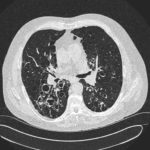
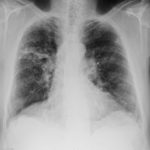
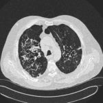
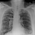
Patients has history of ABPA complicating long standing asthma. His total IgE has fluctuated between 2,200 and 4,600 KU/L, his Aspergillus IgE between 36.3 and 65.4 kAU/L and Aspergillus IgG from 87-154 mg/L. He has been taking long term itraconazole.
 ,
,  ,
,  ,
, 

 ,
,  ,
, 