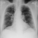
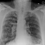
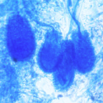
Chronic cavitary invasive aspergillosis in a non-immunocompromised patient, pt RW.

Date: 26 November 2013

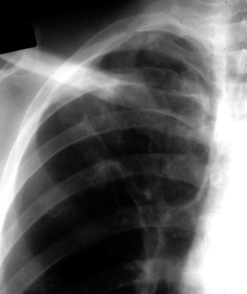
  • Image A. December 1991 Close up view of right upper-lobe of the lung in a 45 year old man who smoked cigarettes showing an ill-defined shadow behind the clavicle and additional abnormalities inferior to this in the right upper-lobe.

    Image A. December 1991 Close up view of right upper-lobe of the lung in a 45 year old man who smoked cigarettes showing an ill-defined shadow behind the clavicle and additional abnormalities inferior to this in the right upper-lobe.

  • Image B. June 1992 Recurrence of disease.Chest radiograph demonstrating cavitary invasive aspergillosis.

    Image B. June 1992 Recurrence of disease.Chest radiograph demonstrating cavitary invasive aspergillosis.

  • Image C. September 1992 Chest radiograph demonstrating further progression of pulmonary aspergillosis with multiple cavities in the right upper-lobe.

    Image C. September 1992 Chest radiograph demonstrating further progression of pulmonary aspergillosis with multiple cavities in the right upper-lobe.

  • Image D. September 2002 Well, although had a respiratory infection. CRP and ESR normal in August 02, aspergillus precipitins positive at a titre of 1:2.

    Image D. September 2002 Well, although had a respiratory infection. CRP and ESR normal in August 02, aspergillus precipitins positive at a titre of 1:2.

  • Image E. January 03 Much worse with lethargy, anorexia, weight loss and radiological deterioration on Chest Xray (link). Aspergillus precipitins rose to a titre of 1:32 , and CRP to 30, and ESR to 49. He was restarted on itraconazole.

    Image E. January 03 Much worse with lethargy, anorexia, weight loss and radiological deterioration on Chest Xray (link). Aspergillus precipitins rose to a titre of 1:32 , and CRP to 30, and ESR to 49. He was restarted on itraconazole.

  • Image F. April 03 No better on itraconazole despite high concentrations of itraconazole in blood.

    Image F. April 03 No better on itraconazole despite high concentrations of itraconazole in blood.

  • Image G. October 03 Stable and reasonable, with weight loss reversed, CRP <5, ESR 13 and Aspergillus precipitins at a titre of 1:16 . Further evolution of radiological features.

    Image G. October 03 Stable and reasonable, with weight loss reversed, CRP <5, ESR 13 and Aspergillus precipitins at a titre of 1:16 . Further evolution of radiological features.

  • Image H. April 04 Back to where he was in terms of symptoms when he stopped itraconzole. CRP, <5, ESR 20, Aspergillus precipitins titre 1:8. Further radiological change.

    Image H. April 04 Back to where he was in terms of symptoms when he stopped itraconzole. CRP, <5, ESR 20, Aspergillus precipitins titre 1:8. Further radiological change.

  • Image I. June 08 Patient remained well no cough, no sputum despite radiological signs.

    Image I. June 08 Patient remained well no cough, no sputum despite radiological signs.

  • Image J. Oct 09 X ray showed litle change over last 16 months in reasonable health, continuing on itraconazole.

    Image J. Oct 09 X ray showed litle change over last 16 months in reasonable health, continuing on itraconazole.

  • Image Image K. A CT scan section of a part of the right upper-lobe lesion shown in 2IPA9.

    Image Image K. A CT scan section of a part of the right upper-lobe lesion shown in 2IPA9.

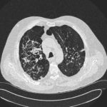
  • Image L. This CT scan of the thorax illustrates the formation of multiple cavities, without aspergillomas previously in ‘normal’ lung a non immunocompromised patient. The appearances are reminiscent of tuberculosis and coccidioidomycosis.

    Image L. This CT scan of the thorax illustrates the formation of multiple cavities, without aspergillomas previously in ‘normal’ lung a non immunocompromised patient. The appearances are reminiscent of tuberculosis and coccidioidomycosis.

  • Image M.

    Image M.

  • Image N.

    Image N.

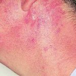
  • Image O. Patient RW: Voriconazole rash. Facial erythema following 9 weeks of oral therapy with voriconazole. The patient noted a sensation of tightness on the skin of his forehead and discomfort in hot water.

    Image O. Patient RW: Voriconazole rash. Facial erythema following 9 weeks of oral therapy with voriconazole. The patient noted a sensation of tightness on the skin of his forehead and discomfort in hot water.

Further details

Image A. December 1991. The lesions were considered to be possibly malignant and surgically resected. Histological examination showed granulomata containing hyphae consistent with Aspergillus. Fungal cultures were not done.

Image B. June 1992 Despite resection of part of the right upper-lobe, invasive aspergillosis recurred six months later. Sputum cultures grew A.fumigatus and Aspergillus antibodies were detected in serum. He responded to itraconazole but subsequently progressed.

Image C. September 1992 Although the appearance suggests the formation of aspergillomas, the contemporaneous CT scan did not confirm this.

Image K. December 1991. There are no particular distinguishing characteristics of invasive aspergillosis. The right upper-lobe was resected and histological examination showed granulomata containing hyphae consistent with Aspergillus. Fungal cultures were not done.

Image M & N. CT scan of thorax. Post RVL segmentectomy with chronic pulmonary aspergillosis. CT shows patchy consolidation with cavitation in the apical segment of right lower lobe. A tiny pneumothorax can be identified posterolaterally (arrows).

Image O. Reference: Muco-cutaneous retinoid effects and facial erythema related to the novel triazole antifungal agent voriconazole. Denning, DW & Griffiths, CEM. Clin.Exp Dermatol 2001, 26(8), 648-53.

Copyright:

Courtesy of Dr D Denning, Wythenshawe Hospital, Manchester.(© Fungal Research Trust)

Notes: n/a


Images library

Showing 10 posts of 2574 posts found.
  • Title

    Legend

  • Drug rashes: Drug interactions between steroids and anti-fungal drugs – (ecchymosis)
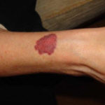
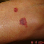
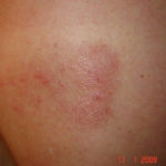
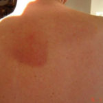
    Image A: Red skin rash on arms due to effect of inhaled steroids termed ecchymosis., Image B: Red skin rash on arms due to effect of inhaled steroids termed ecchymosis., Image C: Papular rash with blisters on the back, associated with CPA but prior to treatment, rash appeared 2 years previously in October 2007. The rash resolved on treatment with azoles., Image D: Papular rash with blisters on the back, associated with CPA but prior to treatment, rash appeared 2 years previously in October 2007. The rash resolved on treatment with azoles.

  • Reference: Muco-cutaneous retinoid effects and facial erythema related to the novel triazole antifungal agent voriconazole. Denning, DW & Griffiths, CEM. Clin.Exp Dermatol 2001, 26(8), 648-53.
    Courtesy of Dr D Denning, Wythenshawe Hospital, Manchester.(© Fungal Research Trust)
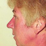
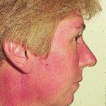
    Patient AB: Voriconazole rash. Following 8 weeks of Voriconazole, patient had remarkable facial erythema, most consistent with sun exposure because of sparing of her neck., Patient AB: Voriconazole rash. Following 8 weeks of Voriconazole, patient had remarkable facial erythema, most consistent with sun exposure because of sparing of her neck., Patient AB: Discoid lupus erythematosus following 12 months voriconazole therapy. This improved with use of sunblock factor 30 and resolved after discontinuation of voriconazole, 2 months later.

  • Micrographs of A. niger conidia & conidial heads provided by Amaliya Stepanova, Head of Laboratory pathomorphology and cytology at Kashkin Research Institute, Russian Federation.
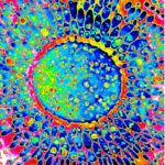
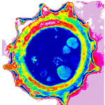
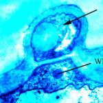
    A niger conidial head TEM in vitro (РКПГF-1124), A niger mature conidium TEM in vitro (РКПГF-1124)

  • Micrographs of A. terreus conidia & conidial heads provided by Amaliya Stepanova, , Head of Laboratory pathomorphology and cytology at Kashkin Research Institute, Russian Federation.
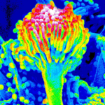
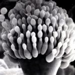
    A terreus aleurospore TEM in vitro (РКПГF-1275), A terreus mature conidium SEM in vitro (РКПГ-1275), A terreus mature conidial head SEM in vitro

  • Micrographs of A. fumigatus conidia & conidial heads provided by Amaliya Stepanova, , Head of Laboratory pathomorphology and cytology at Kashkin Research Institute, Russian Federation.
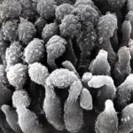
    Conidial head (SEM), Part of conidial head (SEM), Mature conidia (SEM), Hyphae (SEM), Murine lung tissue (TEM)

  • Isolate from environmental sample

    A. clavatus, A. clavatus, A. clavatus

  • Patients has history of ABPA complicating long standing asthma. His total IgE has fluctuated between 2,200 and 4,600 KU/L, his Aspergillus IgE between 36.3 and 65.4 kAU/L and Aspergillus IgG from 87-154 mg/L. He has been taking long term itraconazole.
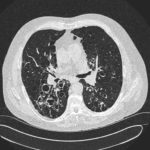
    December 2012, May 2012, AW CT Dec 2012 2, February 2010