Date: 26 November 2013
This recording of peak flow was taken prior to and during the first 4 weeks of inhaled steroids (Becotide 100 and Duovent both 2 puffs 4x daily). The patient had had asthma since age 4, and been treated with bronchodilators and oral courses of steroids when severely affected. The chart, which the patient completed at home, shows that early in week one her peak flow varied from 200-250 L/min. As the medication started to work, the peak flows gradually increased to reach 360-420 L/min in the 4th week. The lower value each morning is characteristic of asthma.
The response to steroids is important confirmation of the diagnosis of asthma (reversible airways obstruction). Many years later she developed ABPA, while on inhaled steroids, with severe upper lobe central bronchiectasis, an IgE of 6,800 Kiu/L, positive aspergillus precipitins, an Aspergillus RAST of 58.7KUa/L (normal <0.4) and eosinophilia.
Copyright: n/a
Notes: n/a
Images library
-
Title
Legend
-
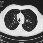
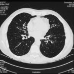
High resolution CT scan images with reconstruction of 1mm thick slices at approximately 10mm increments. The scan shows moderately severe multi-lobar cylindrical and varicose bronchiectasis predominantly centrally and in the upper lungs. There is no mucus plugging seen.
The features are in keeping with allergic bronchopulmonary aspergillosis
 ,
,  ,
, 
-
pt.SB – 6/10/98 – bronchocentric granulomatosis. CT scan showing multiple small nodules of variable size in both lung fields, apparently close to the vascular bundles.

-
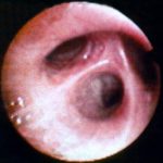
Bronchial oedema.Remarkably oedematous bronchial mucosa, as seen in ABPA.
-
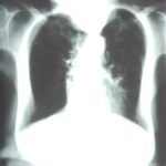
An example of longstanding allergic bronchopulmonary aspergillosis in a patient who has been steroid dependent for over 15 years showing remarkable kyphoscoliosis and honey combing and fibrosis of both lungs.
-
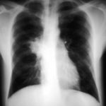
Recurrent pulmonary shadows 1. 6 Jan 1988 – chest radiograph showing right hilar enlargement, consistent with ABPA.
Recurrent pulmonary shadows 1. 3 Feb 1989 – chest radiograph showing right upper-lobe consolidation and contraction consistent with obstruction of RUL bronchus, in ABPA.
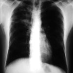
Clearing of pulmonary shadows 3, pt BJ. 5 April 1989 – resolution of shadows seen in February, with a course of corticosteroids.
Recurrence of pulmonary shadows 4, pt BJ. 2 September 1989 – recurrence of pulmonary shadows with an exacerbation of ABPA.
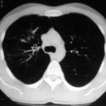
Central bronchiectasis, pt BJ. CT scan of thorax October 1989 showing central bronchiectasis, characteristic of ABPA (and cystic fibrosis).
 ,
,  ,
,  ,
,  ,
, 
-
A typical example of a wet mount of a sputum sample from a patient with allergic bronchopulmonary aspergillosis.


