Date: 26 November 2013
This recording of peak flow was taken prior to and during the first 4 weeks of inhaled steroids (Becotide 100 and Duovent both 2 puffs 4x daily). The patient had had asthma since age 4, and been treated with bronchodilators and oral courses of steroids when severely affected. The chart, which the patient completed at home, shows that early in week one her peak flow varied from 200-250 L/min. As the medication started to work, the peak flows gradually increased to reach 360-420 L/min in the 4th week. The lower value each morning is characteristic of asthma.
The response to steroids is important confirmation of the diagnosis of asthma (reversible airways obstruction). Many years later she developed ABPA, while on inhaled steroids, with severe upper lobe central bronchiectasis, an IgE of 6,800 Kiu/L, positive aspergillus precipitins, an Aspergillus RAST of 58.7KUa/L (normal <0.4) and eosinophilia.
Copyright: n/a
Notes: n/a
Images library
-
Title
Legend
-
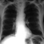
pt FT. Normal chest radiograph of patient with extensive pseudomembranous Aspergillus tracheobronchitis, 4 days before death.
-
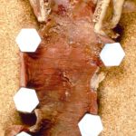
Pt FT. Autopsy appearance of the trachea, after the adherent pseudomembrane had been removed, revealing confluent ulceration superiorly with small green plaques of Aspergillus growth on the trachea inferiorly.
-
This view was obtained in a lung transplant recipient at bronchoscopy. Aspergillus fumigatus was grown from bronchial lavage but invasion was not demonstrated on bronchial biopsy. Symptoms improved with itraconazole therapy and abnormal appearances had resolved within 2 weeks.

-
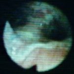
Bronchoscopic view of Aspergillus tracheobronchitis. Bronchial lavage revealed hyphae in microscopy and cultures grew A.fumigatus. This man had received a lung transplant a few weeks before. Invasion of mucosa, but not cartilage, was demonstrated histologically. He responded rapidly to oral itraconazole.
-
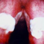
This view from indirect laryngoscopy illustrates bilateral lesions on the larynx that on biopsy were shown to be due to Aspergillus. This is a rare disease in non-immunocompromised patients.
-
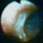
Bronchoscopic view of a deep bronchial ulcer in a lung transplant patient. Biopsies through the ulcer yielded cartilage with hyphae invading it. Fungal cultures of bronchial lavage grew Aspergillus fumigatus. He responded to oral itraconazole therapy.
-
Patient had life threatening pneumonia, cavity formation was later observed. He later presented with a fungal ball. The aspergilloma was removed by surgical resection of the right upper lobe.
 ,
,  ,
,  ,
,  ,
,  ,
,  ,
, 