Date: 26 November 2013
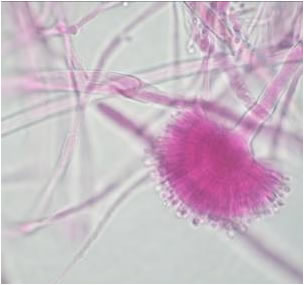
Conidial heads from culture on CYA 25°C medium (mag x100)
Copyright:
These images were generously provided by Mirca Zotti, University of Genoa who retains the Copyright
Notes: n/a
Images library
-
Title
Legend
-
Scanning electron micrograph of Aspergillus ochraceopetaliformis conidial heads

-
Image D & E. A case of onychomycosis associated with Aspergillus ochraceopetaliformis as described in Nail infection by Aspergillus ochraceopetaliformis. Med Mycol. 2009 Mar 9:1-5, 2009, Brasch J, Varga J, Jensen JM, Egberts F & Tintelnot K
 ,
,  ,
,  ,
,  ,
, 
-
Further details
Image 5. Oral itraconazole pulse therapy was given to the patient (200 mg twice daily for 1 week, with 3 weeks off between successive pulses, for four pulses) and treatment was successful.
 ,
,  ,
,  ,
,  ,
, 
-
This patient was 28 yr old with adult lymphocytic leukaemia. She received induction chemotherapy and this infection developed 2 days after recovering from neutropenia.
 ,
,  ,
,  ,
,  ,
,  ,
,  ,
,  ,
,  ,
,  ,
, 
-
Close-up image of the lesion on the left thigh showing a mat of hyphae over the wound.

-
Eosinophilic mucin with A. flavus in the nasal cavity. Irregular crust of 2.5 cm from a patient diagnosed as allergic fungal sinusitis. Patient with allergic fungal sinusitis

-
GMS stain of eosinophilic mucin reveals a darkly stained dichotomously branched A. flavus hyphae within cellular background. Patient with allergic fungal sinusitis


