Date: 7 May 2013
A four day A. fumigatus culture on malt extract agar (above). Light microscopy pictures are taken at 1000x mag., stained with lacto-phenol cotton blue (right).
Copyright:
With thanks to Niall Hamilton.
Notes:
Colonies on CYA 40-60 mm diam, plane or lightly wrinkled, low, dense and velutinous or with a sparse, floccose overgrowth; mycelium inconspicuous, white; conidial heads borne in a continuous, densely packed layer, Greyish Turquoise to Dark Turquoise (24-25E-F5); clear exudate sometimes produced in small amounts; reverse pale or greenish. Colonies on MEA 40-60 mm diam, similar to those on CYA but less dense and with conidia in duller colours (24-25E-F3); reverse uncoloured or greyish. Colonies on G25N less than 10 mm diam, sometimes only germination, of white mycelium. No growth at 5°C. At 37°C, colonies covering the available area, i.e. a whole Petri dish in 2 days from a single point inoculum, of similar appearance to those on CYA at 25°C, but with conidial columns longer and conidia darker, greenish grey to pure grey.
Conidiophores borne from surface hyphae, stipes 200-400 µm long, sometimes sinuous, with colourless, thin, smooth walls, enlarging gradually into pyriform vesicles; vesicles 20-30 µm diam, fertile over half or more of the enlarged area, bearing phialides only, the lateral ones characteristically bent so that the tips are approximately parallel to the stipe axis; phialides crowded, 6-8 µm long; conidia spherical to subspheroidal, 2.5-3.0 µm diam, with finely roughened or spinose walls, forming radiate heads at first, then well defined columns of conidia.
Distinctive features
This distinctive species can be recognised in the unopened Petri dish by its broad, velutinous, bluish colonies bearing characteristic, well defined columns of conidia. Growth at 37°C is exceptionally rapid. Conidial heads are also diagnostic: pyriform vesicles bear crowded phialides which bend to be roughly parallel to the stipe axis. Care should be exercised in handling cultures of this species.
Images library
-
Title
Legend
-
Further image details
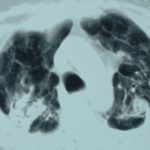
Image A. Long standing sarcoidosis, on corticosteroids with fibrosis and cavitary disease, and a possible fungal ball in the cavity on the left (1996).
Image B. Long standing sarcoidosis, on corticosteroids with 2 cavities containing aspergillomas, one on the left and one on the right (1996).
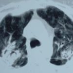
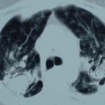
Image C. Sarcoidosis with progressive cavity formation and aspergillomas. Probable CIPA given appearances (2000).
Image D. Sarcoidosis with progressive cavity formation and aspergillomas. Probable CIPA given appearances (2000).
Image E. Sarcoidosis with progressive cavity formation and aspergillomas. Probable CIPA given appearances (2000).
 ,
,  ,
,  ,
,  ,
, 
-
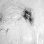
Late (venous) phase angiogram of a right intercostal artery showing persistence of vascular blush and further filling of a branch of the pulmonary artery.
-
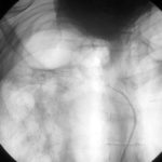
Catheter tip in a right posterior intercostal artery on screening film.
-
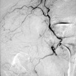
Angiogram of a right bronchial artery on subtraction film in the early arterial phase showing filling hypervascular circulation superiorly and communications with a pulmonary arterial radical.
-
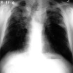
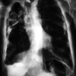
This patient with severe pulmonary sarcoidosis has bilateral aspergillomas. A rim of air is visible around parts of the aspergillomas on both sides. This patient was recruited into the NIAID Mycoses Study Group multicentre study of the treatment of invasive pulmonary aspergillosis with itraconazole but not analysed because invasive disease was not demonstrated. Denning DW, Lee JY, Hostetler JS, Pappas P, Kauffman CA, Dewsnup DH, Galgiani JN, Graybill JR, Sugar AM, Catanzaro A, Gallis H, Perfect JR, Dockery B, Dismukes WE, Stevens DA, NIAID Mycoses Study Group multicenter trial of oral itraconazole therapy of invasive aspergillosis. Am J Med 1994; 97: 135-144
-
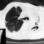
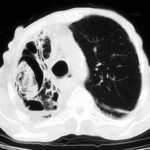
Extensive pleural thickening is demonstrated at the left apex on this CT scan of a woman who had previously had tuberculosis and whose large cavity gradually became obliterated by pleural thickening. An aspergilloma is demonstrable within the cavity
-
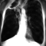
This chest radiograph (AMBER film) demonstrates the typical extensive pleural thickening at the right apex, seen in patients with aspergillomas. The cavity appears not to contain an aspergilloma but on CT scan had some ‘debris’ and Aspergillus antibiotics (precipitins) were strongly positive. The differential diagnosis lies between an aspergilloma and chronic invasive pulmonary aspergillosis. The extensive pleural thickening is heavily in favour of an aspergilloma, even without a well demonstrated fungal ball in the cavity.
-
Image C. Another example of a severe apical aspergilloma with remarkably little pleural thickening on plain chest radiograph (AMBER film). Severe distortion of the trachea is demonstrated.
 ,
,  ,
, 

![Asp[1]fumigatus](https://www.aspergillus.org.uk/wp-content/uploads/2013/11/Asp1fumigatus.jpg)

