Date: 7 May 2013
Copyright: n/a
Notes:
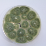
Colonies on CYA 40-60 mm diam, plane or lightly wrinkled, low, dense and velutinous or with a sparse, floccose overgrowth; mycelium inconspicuous, white; conidial heads borne in a continuous, densely packed layer, Greyish Turquoise to Dark Turquoise (24-25E-F5); clear exudate sometimes produced in small amounts; reverse pale or greenish. Colonies on MEA 40-60 mm diam, similar to those on CYA but less dense and with conidia in duller colours (24-25E-F3); reverse uncoloured or greyish. Colonies on G25N less than 10 mm diam, sometimes only germination, of white mycelium. No growth at 5°C. At 37°C, colonies covering the available area, i.e. a whole Petri dish in 2 days from a single point inoculum, of similar appearance to those on CYA at 25°C, but with conidial columns longer and conidia darker, greenish grey to pure grey.
Conidiophores borne from surface hyphae, stipes 200-400 µm long, sometimes sinuous, with colourless, thin, smooth walls, enlarging gradually into pyriform vesicles; vesicles 20-30 µm diam, fertile over half or more of the enlarged area, bearing phialides only, the lateral ones characteristically bent so that the tips are approximately parallel to the stipe axis; phialides crowded, 6-8 µm long; conidia spherical to subspheroidal, 2.5-3.0 µm diam, with finely roughened or spinose walls, forming radiate heads at first, then well defined columns of conidia.
Distinctive features
This distinctive species can be recognised in the unopened Petri dish by its broad, velutinous, bluish colonies bearing characteristic, well defined columns of conidia. Growth at 37°C is exceptionally rapid. Conidial heads are also diagnostic: pyriform vesicles bear crowded phialides which bend to be roughly parallel to the stipe axis. Care should be exercised in handling cultures of this species.
Images library
-
Title
Legend
-
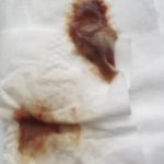
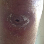
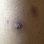
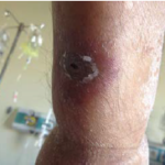
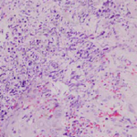
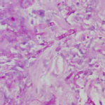
Images and abstract taken from Mert D et.al., Hematol Rep. 2017 Jun 1;9(2):6997. doi: 10.4081/hr.2017.6997. Invasive Aspergillosis with Disseminated Skin Involvement in a Patient with Acute Myeloid Leukemia: A Rare Case.
Invasive pulmonary aspergillosis is most commonly seen in immunocompromised patients. Besides, skin lesions may also develop due to invasive aspergillosis in those patients. A 49-year-old male patient was diagnosed with acute myeloid leukemia.
The patient developed bullous and zosteriform lesions on the skin after the 21st day of hospitalization. The skin biopsy showed hyphae. Disseminated skin aspergillosis was diagnosed to the patient.
Voricanazole treatment was initiated. The patient was discharged once the lesions started to disappear.
 ,
,  ,
,  ,
,  ,
, 
-
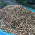
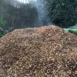
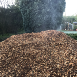
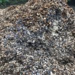
A pile of woodchip stored for use in a garden usually as a weed suppressing mulch. The heat building up in the pile is illustrated by the plumes of steam eminating from the top of the pile.
Aspergillus fumigatus is particularly well adapted to grow in the heat (up to 60C) found in such piles of rotting organic material and this characteristic, an adaption for its life in its natural environment also enables it to survive and grow in warm mammalian bodies at 37C. Most fungi cannot grow or survive at those temperatures
 ,
,  ,
,  ,
, 
-
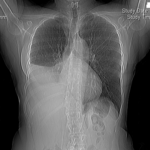
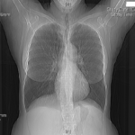
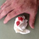
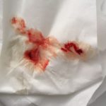
MK is 59 years old and presented with right sided pleuritic chest pain and coughing over 1 week. A chest Xray and then CT scan revealed complete collapse of her right lower lobe and middle lobes. Mucous retention is seen just proximal to the abrupt cutoff. There was mild bronchiectasis.
 ,
, 


 ,
,  ,
, 