Date: 26 November 2013
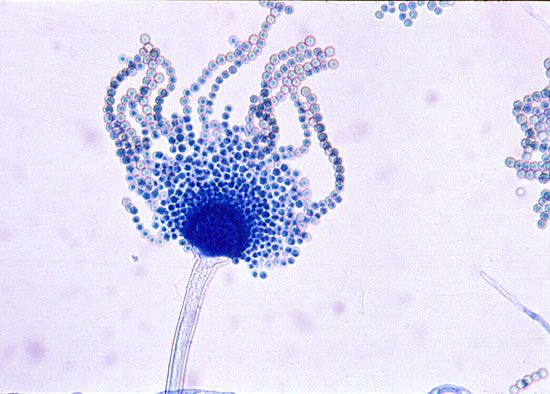
Microscopic morphology of A. flavus. Conidial heads are radiate, splitting to form loose columns, biseriate but having some heads with phialides borne directly on the vesicle.
Copyright:
With thanks to G Kaminski. D Ellis and R Hermanis Mycology Unit Women’s & Children’s Hospital , Adelaide, South Australia 5006. © Fungal Research Trust
Notes:
Colonies on CYA 60-70 mm diam, plane, sparse to moderately dense, velutinous in marginal areas at least, often floccose centrally, sometimes deeply so; mycelium only conspicuous in floccose areas, white; conidial heads usually borne uniformly over the whole colony, but sparse or absent in areas of floccose growth or sclerotial production, characteristically Greyish Green to Olive Yellow (1-2B-E5-7), but sometimes pure Yellow (2-3A7-8), becoming greenish in age; sclerotia produced by about 50% of isolates, at first white, becoming deep reddish brown, density varying from inconspicuous to dominating colony appearance and almost entirely suppressing conidial production; exudate sometimes produced, clear, or reddish brown near sclerotia; reverse uncoloured or brown to reddish brown beneath sclerotia. Colonies on MEA 50-70 mm diam, similar to those on CYA although usually less dense. Colonies on G25N 25-40 mm diam, similar to those on CYA or more deeply floccose and with little conidial production, reverse pale to orange or salmon. No growth at 5°C. At 37°C, colonies usually 55-65 mm diam, similar to those on CYA at 25°C, but more velutinous, with olive conidia, and sometimes with more abundant sclerotia.
Sclerotia produced by some isolates, at first white, rapidly becoming hard and reddish brown to black, spherical, usually 400- 800 µm diam. Teleomorph not known. Conidiophores borne from subsurface or surface hyphae, stipes 400 µm to 1 mm or more long, colourless or pale brown, rough walled; vesicles spherical, 20-45 µm diam, fertile over three quarters of the surface, typically bearing both metulae and phialides, but in some isolates a proportion or even a majority of heads with phialides alone; metulae and phialides of similar size, 7-10 µm long; conidia spherical to subspheroidal, usually 3.5-5.0 µm diam, with relatively thin walls, finely roughened or, rarely, smooth.
Distinctive features
Aspergillus flavus is distinguished by rapid growth at both 25°C and 37°C, and a bright yellow green (or less commonly yellow) conidial colour. A. flavus produces conidia which are rather variable in shape and size, have relatively thin walls, and range from smooth to moderately rough, the majority being finely rough.
Images library
-
Title
Legend
-
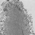
Light microscopic image of hyphae in an aspergilloma (10x magnification)
-
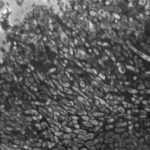
Light microscopic image of hyphae in an aspergilloma (400x magnification)
-
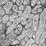
An aspergilloma (or fungal ball) is a mass of fungus found inside the body, for example inside cavities such as the lungs or sinuses, or as abscesses in organs such as the brain or kidney. They are made up of threadlike fungal strands (hyphae) that are densely packed but only around 1/200 of a millimetre in diameter. A mass of hyphae is called a mycelium.
In this image, a slice through an aspergilloma has been imaged using a transmission electron microscope.
-
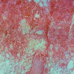
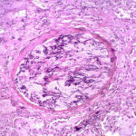
Aspergillus can punch through the lining of the lungs and invade the blood vessels below, in a process called angioinvasion. It can result in blockage (occlusion) of the blood vessel and damage to the local tissue through lack of oxygen (infarction). In severely immunocompromised patients, fragments can even break off and travel to other organs in the body.
In this image, a tissue section through a blocked blood vessel has been stained with the dyes haematoxylin (purple, binds DNA) and eosin (pink, binds proteins).
-
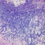
Showing the edge of a colony of aspergillus forming a fungal ball. The fungal hyphae exhibit dichotomous 45 degree angle branching and septae typical of Aspergillus.
-
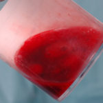
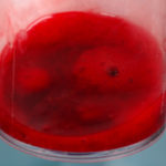
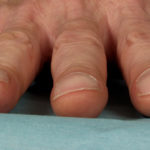
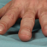
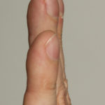
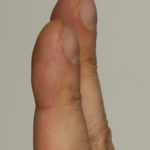
Pt CJ finger clubbing, this patient had chronic cavitary pulmonary aspergillosis, with an aspergilloma since 1988, following an episode of haemoptysis. Currently patient still has symptomatic disease.
Images E,F Blood stained sputum samples from this patient.
 ,
,  ,
,  ,
,  ,
,  ,
, 
-
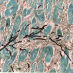
Disseminated, invasive aspergillosis showing dichotomously branching hyphae. Original magnification x300. Stained with Gomori Methenamine Silver (GMS).
-
Disseminated, invasive aspergillosis showing dichotomously branching hyphae. Original magnification x150. Stained with Gomori Methenamine Silver (GMS).

-
Disseminated, invasive aspergillosis showing dichotomously branching hyphae. Original magnification x50. Stained with Gomori Methenamine Silver (GMS).

-
Light microscopical appearance of invasive pulmonary aspergillosis showing vessel occlusion with thrombus and distal infarction (Haematoxylin and eosin, x100)
