Date: 28 July 2023
Copyright:
Arugosins G and H: prenylated polyketides from the marine-derived fungus Emericella nidulans var. acristata. Kralj, Ana; Kehraus, Stefan; Krick, Anja; Eguereva, Ekaterina; Kelter, Gerhard; Maurer, Martina; Wortmann, Andreas; Fiebig, Heinz-Herbert; Koenig, Gabriele M. Journal of Natural Products (2006), 69(7), 995-1000
Notes:
Metabolite structure of Arugosin G
Images library
-
Title
Legend
-
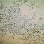
Mucous plug examined by light microscopy with KOH, showing a network of hyaline branching hyphae typical of Aspergillus, from a patient with ABPA.
-
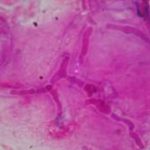
Corneal scraping stained with lactophenol cotton blue showing beaded septate hyphae not typical of either Fusarium spp or Aspergillus spp, being more consistent with a dematiceous (ie brown coloured) fungus
-
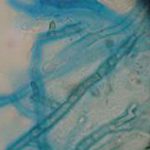
Corneal scrape with lactophenol cotton blue shows separate hyphae with Fusarium spp or Aspergillus spp.
-
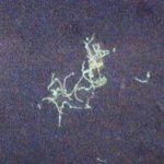
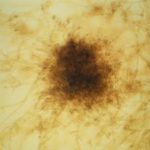
A filamentous fungus in the CSF of a patient with meningitis that grew Candida albicans in culture subsequently.
-
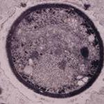
Transmission electron micrograph of a C. neoformans cell seen in CSF in an AIDS patients with remarkably little capsule present. These cells may be mistaken for lymphocytes.
-
India ink preparation of CSF showing multiple yeasts with large capsules, and narrow buds to smaller daughter cells, typical of C. neoformans
-
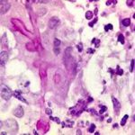
PAS stain. An example of Aspergillus fumigatus.
(PAS-stained) in a patient with chronic granulomatous disease showing a 45 degree branching hypha within a giant cell. Rather bulbous hyphal ends are also seem, which is sometimes found inAspergillus spp. infections, histologically. (x800)


